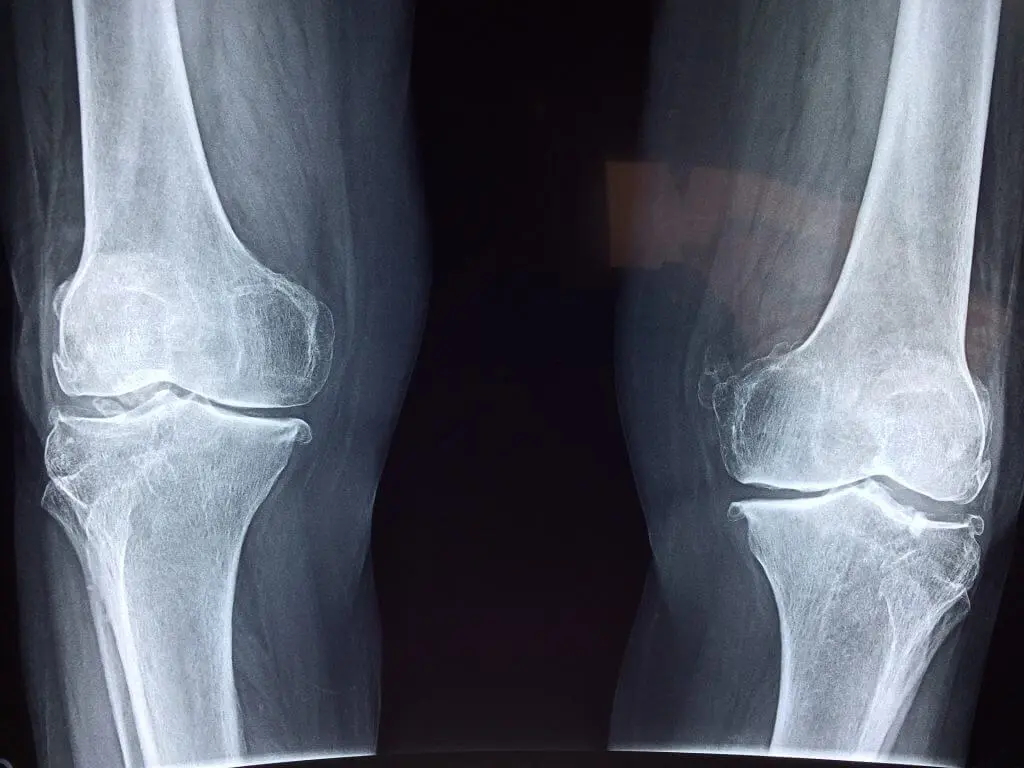
Young people who practise certain sports intensively, but also older people, may at some point feel intense pain at the front of the knee, and the orthopaedic doctor's diagnosis may be: chondropathy or cartilage injury. Let us take a closer look at what this condition involves, what causes it and how it is treated.
What is chondropathy and how many types are there?
First of all, we need to know that every bone end inside the knee joint is covered with cartilage. Its role is both to absorb shocks and to ensure minimal friction between the bones inside the joints. Blows or accidents, degenerative damage, wear and many other factors that we will detail later can injure the cartilage.
People affected by chondropathy will feel pain at the front of the knee, which may sometimes swell, and will have the sensation that the joint is "grinding", as if bone were rubbing against bone. This is the moment when we need to see a doctor urgently.
Chondropathy is divided into four types, depending on the severity of the injury:
- "Fraying"-type lesion
- Superficial fissures
- Deep fissures, less than 75% of the cartilage thickness, with no exposed bone
- Fissures with exposure of the subchondral bone
Risk factors and causes of chondropathy
The risk factors that lead to the development of chondropathy include:
- Young age. During the growth period, temporary musculoskeletal imbalances can occur
- Lower muscle mass and a larger "Q angle" make this condition more common in women than in men
- Some sports – athletics, cycling, rowing, tennis, football – practised intensively are a risk factor
- Knee osteoarthritis (gonarthrosis) favours the development of chondropathy
The common causes of chondropathy include:
- Major trauma (sports or road accidents)
- Wear of the joint in athletes
- Degeneration of the joint in older people (patellofemoral osteoarthritis)
- Hyperlaxity of the knee ligaments
Symptoms of cartilage injury
The most important symptom of chondropathy or cartilage injury is pain. But, in addition to this, there are other troublesome manifestations: hydrarthrosis (fluid on the knee), inflammation, locking of the joint and crepitus (cracking of the bones).
Treatment of chondropathy
There are two types of treatment for chondropathy: conservative and surgical. Depending on the patient's clinical context, the orthopaedic doctor is the one who will decide which of the two is the most appropriate to apply.
Conservative treatment
This is the treatment preferred by both the doctor and the patient, and it can be applied when the symptoms are moderate. Its role is to relieve the symptoms and restore the mobility of the joint. Conservative treatment involves:
- Rest
- Applying ice packs several times a day (but not directly on the skin)
- Stabilising the kneecap with medical devices recommended by the orthopaedic doctor
- Elevating the affected limb above the level of the heart
- Prescribing anti-inflammatory and pain-relieving medication
Surgical treatment of chondropathy
In some more complex cases, involving deep fissures or anatomical defects, the orthopaedic doctor may recommend surgical treatment. The type of surgical treatment is decided according to the pathology of each case.
Types of surgical treatment
For grade 1 and 2 lesions, arthroscopic surgical treatment is used, in which the chondral lesions are stabilised mechanically and thermally, so that the synovial fluid can no longer penetrate the fissures in the cartilage.
For grade 2-4 cartilage lesions, chondral reconstruction is needed. If the affected area is smaller than 1 cm², the microfracture technique is used. This means that small holes are made in the bone left without cartilage, down to the layer containing haematopoietic bone marrow cells. The blood that comes from these holes forms a fibrin network that fills the chondral defect, into which chondrocytes from the adjacent healthy cartilage will migrate over the following weeks. This forms a cartilage of inferior quality compared with normal cartilage, but with very good clinical results. This is a medical procedure that is carried out in a single stage.
If the affected area is larger than 1 cm² – 1.5 cm², other reconstruction techniques are recommended:
- Chondral matrix implantation (AMIC)
- Mosaicplasty
- Autologous chondrocyte implantation (ACI)
The technique preferred by Dr. Ion Bogdan Codorean for chondral reconstruction in grade 3-4 chondropathies with an area of over 1.5 cm² is chondral matrix implantation (scaffold). The chondrocytes migrate into this matrix, with the aim of transforming into hyaline cartilage of superior quality to the pseudo-cartilage generated by microfractures. Depending on the size of the chondral lesion and its location, the chondral implant is performed arthroscopically or through a mini-arthrotomy.
Chondral matrix implantation (AMIC)
This is also a two-stage surgical technique, the difference from ACI being that the chondral cells are cultured on a three-dimensional matrix. The matrix is attached with fibrin glue or sutured. Chondral matrix implantation is a technique frequently used for large cartilage lesions.
Mosaicplasty
This consists of harvesting autologous cartilage together with a bone cylinder from an area of minimal importance in the knee, which is then implanted into the area of damaged cartilage. After this stage, the chondrocytes remain viable and the bone graft incorporates. Mosaicplasty can be performed arthroscopically, in a single stage. This technique has the disadvantage that it requires harvesting from healthy areas of the knee, with the possibility of pain developing at the harvest site.
Autologous chondrocyte implantation (ACI)
This is a treatment in two surgical stages. In the first stage, chondral tissue is harvested from an area of minimal importance, followed by a period in which the chondrocytes are cultured in the laboratory. In the second surgical stage, the cartilage obtained by culturing the patient's own chondral cells is implanted.
Rehabilitation after surgery
Healing and rehabilitation after surgery comprise several stages and will be adapted to the individual condition of the patient. In the first phase, during the first two weeks after the operation, the aims are: protecting the patellar tendon, reducing local oedema, promoting healing, achieving 90 degrees of flexion, achieving and maintaining full extension, and muscle strengthening. The orthopaedic surgeon may recommend wearing compression stockings (to prevent the formation of blood clots), a crutch or brace, applying ice packs and sets of rehabilitation exercises.
Over the following two to four weeks, the focus will be on regaining full mobility of the knee and beginning muscle toning, with the rehabilitation process after chondropathy surgery expected to be completed within twelve weeks.

Dr. Ion Bogdan Codorean
Senior consultant in orthopaedics and sports traumatology. PhD in medicine.
View profileNewsletter subscription
Sign up for the Artro Sport newsletter free of charge!